Cerebral malaria, a severe form of malaria, affects millions of people worldwide. While the central nervous system is the primary target, the eye can also be impacted by this debilitating condition. Malarial retinopathy, a unique retinal manifestation of cerebral malaria, can provide valuable insights into the pathophysiology and severity of the disease. In this blog, we will explore the intricate connection between cerebral malaria and the eye, understanding the mechanisms behind malarial retinopathy, its implications, and possible interventions.
The Link between Cerebral Malaria and the Eye:
Cerebral malaria is caused by the Plasmodium falciparum parasite, transmitted by mosquito bites. As it infects red blood cells, it can result in severe systemic complications. The retina, being an extension of the central nervous system, becomes vulnerable to the effects of the infection. Malarial retinopathy occurs when the blood vessels in the retina are affected, leading to various structural and functional changes.
- Clinical Features of Malarial Retinopathy:
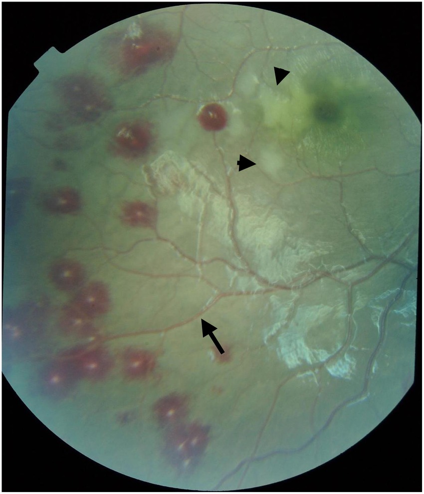
Malarial retinopathy exhibits specific clinical features that can aid in the diagnosis and assessment of cerebral malaria. These include retinal whitening, vessel changes, hemorrhages, and cotton wool spots, among others. Understanding these features and their significance can help in predicting disease severity and monitoring response to treatment. - Pathophysiology and Mechanisms:
The precise mechanism behind malarial retinopathy is not yet fully understood. However, several hypotheses have been proposed. It is believed that the parasite-induced inflammation and immune response trigger endothelial dysfunction and disruption of the blood-retinal barrier. This, in turn, leads to retinal vascular pathology and subsequent retinal changes. - Implications for Disease Severity and Prognosis:
Malarial retinopathy has demonstrated strong associations with the severity of cerebral malaria and poor clinical outcomes. The extent of retinal involvement, including the presence of specific retinal signs, is correlated with increased mortality rates, coma duration, and neurological sequelae. Thus, assessing malarial retinopathy can provide valuable prognostic information to guide treatment decisions. - Role in Monitoring Treatment Response:
Monitoring changes in malarial retinopathy during and after treatment can serve as a non-invasive tool for evaluating the efficacy of antimalarial therapies. Improved retinal findings and resolution of retinal signs correspond with successful treatment and decreased disease severity. - Diagnostic Challenges and Advancements:
Diagnosing malarial retinopathy can be challenging, especially in resource-limited settings. Ophthalmic examination techniques, including direct and indirect ophthalmoscopy, can help identify retinal abnormalities. However, the development of imaging technologies, such as retinal photography and optical coherence tomography (OCT), has revolutionized the diagnosis and monitoring of malarial retinopathy. - Potential Interventions and Research:
Given the crucial role of malarial retinopathy in understanding cerebral malaria, ongoing research aims to investigate potential interventions and therapeutic targets. Strategies such as anti-inflammatory hydrogen therapy, vision therapy and for mitigating retinal damage and improving vision of patient outcomes.
Malarial retinopathy provides profound insights into the pathophysiology, severity, and prognosis of cerebral malaria. By understanding the ocular manifestations of this devastating condition, healthcare professionals can enhance the diagnosis, monitoring, and treatment of cerebral malaria patients. Further research, advancements in imaging techniques, and targeted interventions hold immense potential to mitigate the impact of malarial retinopathy and improve patient outcomes in the fight against cerebral malaria.